Wetenschap
Het netvlies onthullen:de hoornvliescontactlens van grafeen biedt robuuste, irritatievrije topografische elektroretinografie
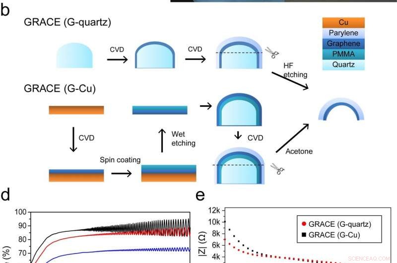
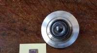
figuur 1 Fabricage en karakterisering van GRACE-apparaten. een Schematische tekening van ERG-opname met het GRACE-apparaat. B Schematische illustratie van GRACE-fabricage met G-kwarts en G-Cu. C Foto's van een GRACE-apparaat gemaakt van G-kwarts. Schaalbalk, 3mm. Afbeelding in de inzet toont de hoge zachtheid van het GRACE-apparaat. NS Optische transmissie van de kale Parylene-C, en GRACE-apparaten gemaakt van G-kwarts en G-Cu, allemaal met een Paryleen-dikte van 25 m. De transmissie bij een golflengte van 550 nm wordt weergegeven in de inzet. e Omvang en fase van elektrochemische impedantie van GRACE-apparaten gemeten in 1 × PBS (pH 7,4). Krediet:Rongkang Yin, Zheng Xu, Xiaojie Duan, et al. Zachte transparante grafeen contactlenselektroden voor conforme volledige cornea-opname van elektroretinogram. Natuur Communicatie deel 9, Artikelnummer:2334 (2018). Gelicentieerd onder een Creative Commons Attribution 4.0 International-licentie (https://creativecommons.org/licenses/by/4.0/legalcode).
Ons zicht kan worden beschadigd of verloren gaan door schade aan het netvlies - een sensorisch membraan aan de achterkant van het oog dat licht waarneemt, het gevormde beeld omzetten in elektrochemische neuronale signalen - resulterend uit twee klassen medische aandoeningen:een aantal erfelijke degeneratieve aandoeningen - waaronder retinitis pigmentosa, aangeboren amaurose van Leber, kegeldystrofie, en Ushersyndroom, evenals diabetische retinopathie, centrale retinale aderocclusie, sikkelcelretinopathie, toxische en auto-immuunretinopathieën, netvliesloslating, en andere oogaandoeningen. Om de juiste diagnose en behandeling te krijgen (vooral wanneer cataract oftalmoscopie in gevaar brengt, 2D fundusfotografie, 3D optische coherentietomografie, en andere hulpmiddelen voor retinale beeldvorming), dergelijke medische aandoeningen zijn afhankelijk van elektroretinografie - een gevoelige techniek die elektrische potentiaalveranderingen op het hoornvliesoppervlak van het oog detecteert en meet, geproduceerd als reactie op een lichtstimulus door neuronale en niet-neuronale retinale cellen. Hoe dan ook, elektroretinografie heeft in het verleden te maken gehad met uitdagingen in de oculaire interface-elektroden die nodig zijn om een elektroretinogram (ERG) te detecteren, dit zijn ongemak voor de patiënt door harde elektroden, beperkte soorten elektroretinogrammen met een enkel type elektrode, verminderde signaalamplitudes en stabiliteit, en overmatige oogbewegingen. Onlangs, echter, wetenschappers aan de Universiteit van Peking, Peking, zacht hebben aangetoond, transparante GRAphene contactlens-elektroden (GRACE's) voor conforme full-cornea elektroretinogram-signaalregistratie bij konijnen en cynomolgus-apen, waaruit blijkt dat hun zachte grafeen-contactlenselektroden deze beperkingen aanpakken.
Prof. Xiaojie Duan besprak het artikel dat zij, afgestudeerde studenten en hoofdauteurs Rongkang Yin en Zheng Xu, en hun co-auteurs gepubliceerd in Natuurcommunicatie . De grootste uitdaging bij het vervaardigen van zachte grafeen contactlenselektroden met breedspectrum optische transparantie, Dr. Duan vertelde: Phys.org , maakte kreukvrije contactlenselektroden, uitleggen dat rimpels optische inhomogeniteit over de elektrode kunnen veroorzaken, waardoor de oculaire breking en de nauwkeurigheid van het lichtstimuluspatroon op het netvlies worden beïnvloed. "Dit ondermijnt op zijn beurt de werkzaamheid van de diagnose van retinopathie, " Dr. Duan voegde toe. "Grafeen verkregen uit de conventionele groeimethode is een vlakke film, en rimpels vormen onvermijdelijk na het overbrengen van de platte grafeenfilm op het gebogen oppervlak. Om een grafeen contactlenselektrode te maken met een hoge elektrische geleidbaarheid en optische uniformiteit over de elektrode, het is belangrijk om direct een gebogen grafeenfilm met uniforme dikte te gebruiken."
Het toepassen van GRACE's op conforme elektroretinografische opname van het volledige hoornvlies vormde geen grote obstakels, zij ging door. "Hoewel er geen primaire moeilijkheid is om GRACE's toe te passen op conforme en volledige cornea-elektroretinografische opnames, zolang de gefabriceerde GRACE's een redelijke impedantie en optische transparantie hebben, we kunnen altijd hoogwaardige ffERG- en mfERG-signalen opnemen. Daarom, om GRACES te krijgen met een redelijke impedantie en optische transparantie, grafeenfilm met velweerstand" - een maat voor de weerstand van dunne films die nominaal uniform van dikte zijn - "onder 2000 Ω / sq en optische transparantie boven 70% zal goed genoeg zijn."
Echter, de belangrijkste uitdaging voor algemene ERG-opname is het meten van multifocale ERG (mfERG) - die tegelijkertijd lokale retinale reacties meet van maximaal 250 retinale locaties binnen de centrale 30 graden topografisch in kaart gebracht - weerspiegelt de retinale respons op stimulatie op een specifiek klein netvliesgebied. "Voor multifocale ERG-metingen, "Dr. Duan vertelde" Phys.org , "het lichtstimulatiepatroon wordt op het netvlies geprojecteerd. Het is daarom belangrijk dat het oog een goede breking heeft, zodat het stimuluspatroon duidelijk kan worden geprojecteerd." In aanvulling, de signaalamplitude van multifocale ERG is slechts ongeveer 1/1000ste van die van conventionele full-field ERG (ffERG, die ERG meet onder volledige retina-stimulatie met een lichtbron onder scotopische (donker-aangepaste) of fotopische (licht-aangepaste) retinale aanpassing), terwijl mfERG een relatief langere opnameperiode vereist, waardoor de gevoeligheid comfort, en stabiele interface met het oog, zeer kritisch voor multifocale ERG-opname. "Conventionele contactlenselektroden hebben de neiging de breking van het oog te veranderen, " merkte ze op, "waardoor ze ongeschikt zijn voor multifocale ERG-opname." Dat gezegd hebbende, andere elektroden (bijv. DTL-elektroden), zal de breking van het oog niet veranderen, maar lijden aan een lage meetgevoeligheid en signaalstabiliteit.
Een andere overweging, Dr. Duan merkte op, is dat de ruimtelijke verdeling van het ERG-potentieel over het hoornvlies al lang een vraag is. "Conventionele elektroden gebruiken ondoorzichtig metaal als opname-elementen, die alleen aan de periferie van het hoornvlies kan worden gelokaliseerd om te voorkomen dat het zicht wordt geblokkeerd - een situatie die ruimtelijk opgeloste ERG-opname op meerdere locaties verhindert, wat nodig is om de ERG-potentiaalverdeling over het hoornvlies te onthullen. Een andere factor is dat voor een conventionele stijve elektrode, er is altijd een dikke traanfilm tussen de elektroden en het hoornvlies, die het potentiaalverschil tussen verschillende locaties op het hoornvlies kan overbruggen." Dit laatste nummer introduceert nog een andere belangrijke uitdaging bij het ophelderen van de ruimtelijke verdeling van ERG over het hoornvlies.
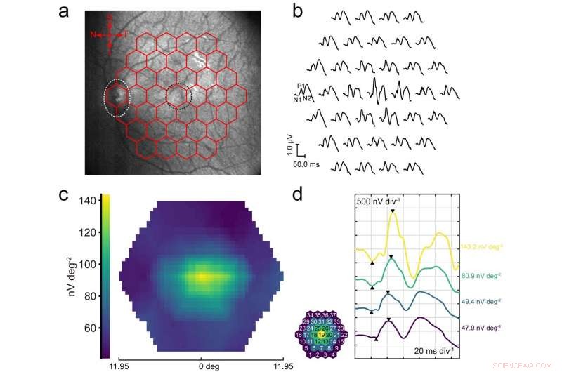
Afb. 3 Multifocale ERG-opname. een Infrarood fundusfoto van een cynomolgus-aapoog genomen tijdens mfERG-opname met een GRACE-apparaat, gesuperponeerd met de stimulus-array. Het wit gestippelde ovaal markeert de positie van de oogzenuwkop en de zwarte gestippelde cirkel markeert de positie van de macula. B Vertegenwoordiging van sporenarray opgenomen vanuit het oog van de cynomolgus-aap in een met GENADE. De golven van 37 focale ERG-signalen zijn topografisch gerangschikt. De belangrijkste mfERG-componenten N1, P1, en N2 kan duidelijk worden gedefinieerd in deze golfvormen, zoals aangegeven bij een van de antwoorden. C Responsdichtheidsplot (retinale weergave) op P1-golf geassocieerd met B . NS De mfERG-antwoorden zijn gegroepeerd en gemiddeld voor elk van de regio's die zijn gemarkeerd met verschillende kleuren. De waarden tonen de responsdichtheid van de P1-piek (zoals gedefinieerd door de driehoeken op de sporen) in elk van de bijbehorende regio's. Krediet:Rongkang Yin, Zheng Xu, Xiaojie Duan, et al. Zachte transparante grafeen contactlenselektroden voor conforme volledige cornea-opname van elektroretinogram. Natuur Communicatie deel 9, Article number:2334 (2018). Licensed under a Creative Commons Attribution 4.0 International License (https://creativecommons.org/licenses/by/4.0/legalcode).
Dr. Duan described the key insights, innovations and techniques they leveraged to address these challenges. "As I mentioned, we eliminate wrinkles by using a curved graphene film directly grown on curved quartz mold—and the film's shape and curvature can be easily tuned by changing those of the quartz molds." The key point, she emphasizes, is the curved graphene film's uniform thickness leads to the resulting GRACEs having uniform electrical conductivity and optical transparency across the entire contact lens electrode, which is what is unique about the team's GRACEs when compared to previously-reported graphene-based eye interfacing devices. "In aanvulling, " she added, "we established and optimized the electrode fabrication flow." She emphasizes that by directly depositing ultrathin insulating film (Parylene-C, which forms the GRACE substrate) onto the graphene/quartz complex, and then etching the quartz mold, GRACE devices can be readily fabricated." The key takeaway is that this fabrication strategy avoids poly(methyl methacrylate) (PMMA)—a transparent thermoplastic (also referred to as an acrylic or acrylic glass) commonly used for graphene transfer, which not only avoids possible PMMA contamination that could cause optical inhomogeneity, but also maintains graphene film integrity—a factor critical to maintaining GRACE electrical conductivity.
As previously noted, it is challenging to record multifocal ERG signals with contact lens electrodes because it tends to alter ocular refraction. "To solve this problem, " Dr. Duan pointed out, "we designed the GRACE to be soft and conformable to the cornea surface with a tight GRACE/cornea interface." This avoids the formation of thick liquid gaps or air gaps between the electrode and the cornea—the main origin of refraction change when wearing hard contact lens electrodes. As shown in their paper, GRACEs can successfully record high-quality multifocal ERG signals, which is indicative of the advantages of GRACEs over hard contact lens electrodes.
To provide efficient multi-site, spatially-resolved ERG recording, the scientists designed and deployed a soft, transparent graphene multi-electrode array. "The soft electrode's tight interface with the cornea avoided tear film shunting, " she explained, "and high optical transparency enables placement of high-density electrode array across the entire corneal surface without blocking the vision or affecting the light stimulus uniformity." Als resultaat, they observed a stronger signal at the central cornea than the periphery, proving the advantages of the soft transparent graphene-based electrodes in ERG recordings.
As to implications of their findings regarding GRACE for in vivo visual electrophysiology studies, Dr. Duan reiterated that their graphene-based contact lens electrodes show the capability for high-efficacy recording of various kinds of ERG recording, including ffERG, mfERG, and meERG (multi-electrode ERG, which maps spatial differences in retinal activity using a conventional full-field stimulus and an array of electrodes on the cornea)—a flexibility not achievable by conventional ERG electrodes. "With further testing and development, " she underscored, "it could replace the traditional electrodes and be used in clinical practice. In addition, because retinal lesions can cause change of the local corneal potentials, the multi-electrode ERG recording with the graphene microelectrode array demonstrated herein provides a potential functional retinal electrophysiological imaging technique that can be used as a diagnostic tool for detecting local areas of retinal dysfunction under single full-field stimulus."
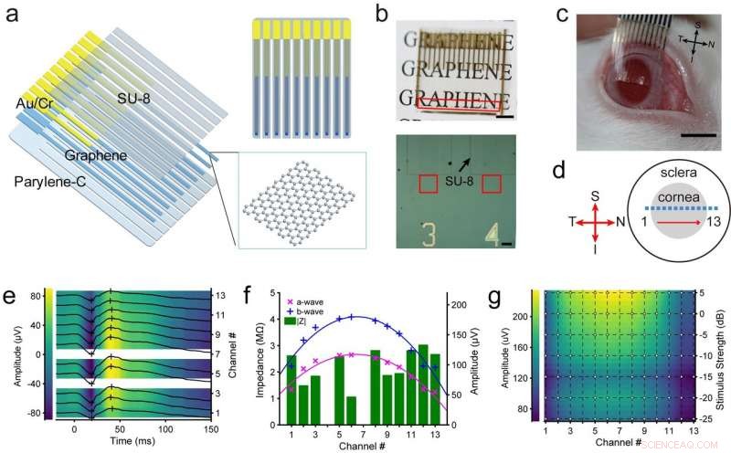
Fig. 5 . Multi-electrode ERG recording with soft, transparent graphene electrode array. een Diagram of graphene multi-electrode array construction showing the layered structures. B Top, a soft, transparent graphene electrode array positioned over a printed paper to show its optical transparency. Scale bar, 5 mm. The recording sites, arranged in a linear pattern, are located in the region marked by the red box. Under each recording site, there is a channel number patterned with Au which is optically opaque. Onderkant, optical microscopy image showing some of the graphene electrode sites and traces. The red box marks the graphene recording sites. The black arrow points to the patterned SU-8 insulation layer on one electrode. Scale bar, 150 μm. C A stripped graphene electrode array positioned over a dilated rabbit eye. Scale bar, 5 mm. NS A schematic drawing showing the positions of the recording channels (marked by the squares) on a rabbit eye. Channel 1 to 13 was evenly distributed over equator of the cornea from temporal to nasal periphery. e A representative set of the multi-electrode scotopic ERG response waveforms. Stimulus strength, 0.3 cd sm−2. The placement of the graphene electrode array is shown in NS . The crosses mark the positions of the a and b- waves. Channels 4 and 7 have abnormally high impedance and are considered non-functional. F Plots of the electrode impedance values |Z| at 100 Hz, a- and b-wave amplitudes of the ERG signals recorded from different channels associated with e . The lines show the quadratic curve fitting of the a- and b-wave amplitudes. G Spatial profile of b-wave amplitudes under different stimulation strength. 0 dB corresponds to 3.0 cd sm−2. The dots in the overlaid grid mark the positions with actual experimental data. Credit:Rongkang Yin, Zheng Xu, Xiaojie Duan, et al. Soft transparent graphene contact lens electrodes for conformal full-cornea recording of electroretinogram. Natuur Communications volume 9, Article number:2334 (2018). Licensed under a Creative Commons Attribution 4.0 International License (https://creativecommons.org/licenses/by/4.0/legalcode).
Moving forward, Dr. Duan identified three planned next steps in the scientists' research, these being:
- Improving electrode gas permeability to make it more suitable for long-term wear
- Fabricate high-density two-dimensional soft transparent electrode array to map the ERG potential across the entire corneal surface
- Apply the soft transparent graphene microelectrode array for in vivo recording of electrical activity of retinal ganglion cells at single-cell level
She also discussed research and other innovations they might consider developing. "Based on nanomaterials and nanotechnology, we seek to develop techniques that can record or modulate neural activities at large scales with high spatio-temporal resolution and long-term stability, and to explore the application of these techniques in understanding fundamental and pathological brain processes."
Tot slot, Dr. Duan listed other areas of research that might benefit from their study. "Soft transparent electrodes also enable simultaneous electrophysiology and optical neural imaging or stimulation, which is important for studying the connectivity and function of neural circuits. Conventional neural surface electrode arrays using opaque metal conductors are not suitable for use in simultaneous electrical and optical neural interfacing because they block the field of view and are prone to producing light-induced artifacts in the electrical recordings. The soft transparent graphene microelectrode array described herein can be used in research combining optical and electrical modalities in neural interfacing."
© 2018 Fys.org
 De wereld wordt geconfronteerd met een wereldwijde zandcrisis
- Wetenschappers helpen de Costa Ricaanse gemeenschap bij het beheren van de slinkende watervoorziening
- Statistieken plot vervuiling om het beleid te informeren
- Levende koraalbedekking zal toekomstige rifoplossing vertragen
- Vergelijk bloeiende planten en coniferen
Hoofdlijnen
- Genomics onthult hoe concurrentie tussen bacteriën de impact van vaccinatie beïnvloedt
- Wetenschappers visualiseren de structuur van de belangrijkste DNA-reparatiecomponent met een bijna-atomaire resolutie
- Lake Michigan watervogels botulisme sterfgevallen in verband met warm water, algen
- Trappers vragen rechtbank om rechtszaak over Amerikaanse bontexport weg te gooien
- Wetenschappers volgen haaien die DNA-fragmenten uit de zee plukken
- Huismuizen kunnen hun vocalisaties moduleren, afhankelijk van het geslacht van de ontvanger
- Nieuwe sorghumcultivars kunnen duizenden liters ethanol produceren
- Team publiceert onderzoek naar ongewone genevolutie in bacteriën
- Basiscelfuncties
- Een nieuwe benadering voor het ontwerpen van de materialen van de toekomst
- Geen goede trillingen meer nodig voor luidsprekers, omdat onderzoek grafeen aanmoedigt om te praten
- Hangende lagen vormen een speciale supergeleider
- Profiteren van grafeendefecten
- 's Werelds eerste commerciële nanogestructureerde bulkmetaal

 Kwam Eiwit, DNA of RNA als eerste?
 - Leraarevaluaties verwijderen slecht presterende leraren in stedelijke scholen
- Onderzoek suggereert dat ijs op de zuidpool van de maan meer dan één bron kan hebben
- Apollo 11-maanmissie trok ruimtegerelateerde bedrijven naar Centraal Florida
- Hulpmiddel om recidive bij federale gevangenen te voorspellen kan ervoor zorgen dat meer gevangenen in aanmerking komen voor vervroegde vrijlating
- Kenmerken van vaatplanten
- Stof zweeft boven het maanoppervlak - elektrostatisch stoftransport hervormt oppervlakken van luchtloze planetaire lichamen
- Langzaamst draaiende radiopulsar gedetecteerd door astronomen
- Elektronica
- Biologie
- Zonsverduistering
- Wiskunde
- French | Italian | Portuguese | Swedish | Dutch | Danish | Norway | Spanish | German |

-
Wetenschap © https://nl.scienceaq.com
